
What is Contractile Force Screening?
Contractile Force Screening (CFS) bypasses molecular intermediates and other indirect endpoints used in current screening technologies, and instead leaps directly to the key physiological endpoint – cellular contractility.
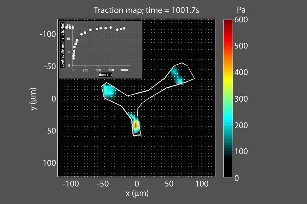
Formerly, cell contractility could be measured by a benchtop technology known as traction force microscopy (TFM), which quantifies the forces that cells exert on their surrounding environment. The principle is simple: If you place a cell on a substrate with a known stiffness, you can measure the deformation the cell exacts on that substrate, and then calculate the traction forces. While TFM is an extremely informative biophysical metric for characterizing changes in cell contractility, its adoption is often limited in biological and biomedical studies to collaborations with researchers in the physical sciences and engineering. This is due to challenges formerly requiring significant expertise and specialized workflows.
Live Cell Technologies Canada bridges this gap by translating TFM from a specialized bench technology into an accessible high-throughput methodology called Contractile Force Screening (CFS). This is done by providing our customers with the four critical customizable elements required for CFS:
Physiological range of stiffness
We have developed formulations of polydimethylsiloxane (PDMS) rubber that are compliant and mechanically tunable, yielding Young's elastic moduli from ~1 kPa to 100 kPa, placing it perfectly within the range of physiological and pathological tissue stiffness.
Fiduciary Fluorescent Beads
We synthesize fluorescent nanoparticle beads and coat them on the substrate surface to visualize cell substrate deformations. No matter the fluorescent labels used in your experiments, we can synthesize beads across the spectrum to fit your imaging needs.
Protein Coatings
We recapitulate the adhesion biochemistry by coating our soft substrates with custom adhesive proteins according to your cell requirements, allowing cells to attach, spread, and generate force on our patented soft substrates.
Software
Finally, we provide analysis software allowing you to extract key contractility metrics from your cell images. Our software dynamically generates and exports data visualizations of these metrics against the dimensions that you select – such as reagent concentration, treatment duration, or cell phenotype – so you can succinctly summarize your insights.
By packing all of this all together into a multi-well format, we put the power of CFS into the hands of the user to explore a number of conditions affecting cell force within a single experiment. Visit our applications page to see how CFS has already been applied in a number of academic studies.